
The term “x-ray” refers to the process of using a beam of energy with a wavelength of 0.01 to 10 nanometers to produce detailed images of the internal structures of the body. This technology is used for a variety of medical applications, including fracture detection. While X-rays can cause a few minor side effects, most adults are safe from them.
X-rays are short energy beams with wavelength in ranges of 0.01 to 10 nanometers
The X-ray is an electromagnetic radiation that has a wavelength of around 0.01 to 10 nanometers. It was, discovered by Wilhelm Conrad Roentgen in 1895 and used to show the content of objects. They can also use to look inside the human body.
X-rays have a wavelength in the range of 0.01 to 10 nanometers, which is in the mid-infrared region of the electromagnetic spectrum. They are not visible to the human eye and are, produced by accelerating charged particles (X-ray tube or synchrotron particle accelerator). Distinct X-rays are, also produced by highly excited atoms. The X-ray region of the electromagnetic spectrum falls outside of visible wavelengths and can detect using photographic film or other types of detectors. This type of imaging is a valuable medical tool.
Monochromatic X-rays are short beams of energy with a wavelength of 0.01 to 10 nanometers. They can use to study various physical, chemical, and mechanical processes. They are also effective in imaging blood vessels and tissues.
X-rays are the most common form of electromagnetic radiation, composed of electrons and are essentially waves of electric and magnetic fields that travel at the speed of light. The electrons that are released from the cathode filament form an electron cloud on the filament’s surface. However, these electrons are not ejected, because they are repelled by the negative charge in the electron cloud.
X-rays are, commonly used for medical purposes, including dental cavities, analysis of astronomical objects, and identifying weld flaws. In addition, X-rays can be used to restore ancient artwork. Despite their usefulness, X-rays can be dangerous to the growing human body, particularly for unborn children. Therefore, pregnant women should always consult a doctor before opting for an X-ray. A doctor will recommend an alternative imaging technique to help reduce the risks to the unborn baby.
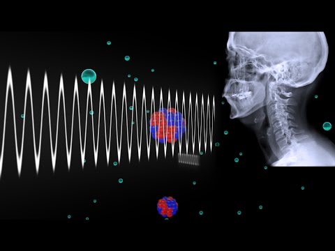
They create detailed images of the structures inside your body
X rays are an important part of a doctor’s diagnostic arsenal. These images can tell doctors about problems ranging from blocked blood vessels to bone cancer. They also help determine whether a particular area of the body is healthy or has some problem, like an infection.
X rays are quick and inexpensive ways to obtain images of the inside of your body. They create detailed images of the structures inside your body by passing invisible x-ray beams through the body. Different materials absorb these beams differently. Dense materials show up as white, while thin or transparent materials appear as black. Bone is the most dense material and is white on an X-ray, while muscle and fat are grey. X-rays are, used to diagnose fractures and other conditions as well as monitor the healing process after surgery.
An x-ray machine sends electromagnetic waves through the body and exposes a thin film that reflects the internal structure. Although x-ray radiation is not harmful, it is important to note that pregnant women and those who are allergic to certain chemicals should avoid having an x-ray.
X-rays can perform on a patient of any age, but women should always tell their providers if they are pregnant or breast-feeding. The radiation from X-rays can harm the fetus. An X-ray can help diagnose symptoms of many illnesses, including cancer and structural issues in bones and joints. It can also be used for routine screenings for cancer.
Used to check for fractures
X rays are diagnostic tools that allow doctors to examine any part of a patient’s body. They use a small amount of radiation to produce images of bones and other soft tissues. These images are very helpful for detecting fractures and broken bones. They can also show the location and type of fracture.
While X rays are the most commonly, used imaging technique for fracture diagnosis, ultrasound is an alternative method for diagnosis. It can identify fractures in an outpatient setting and can save time, money, and resources. It also has high sensitivity, which is important for diagnosing a fracture.
If you think that you may have a fracture, your doctor will perform an X-ray to determine the type and severity. While minor fractures don’t require emergency care, if you are concerned about a serious fracture, you should immediately call 911 for medical attention.
In addition to bone fractures, X-rays can detect bone thinning and osteoporosis. They can also detect lung infections and bone tumors. Chest X-rays also used to monitor lung conditions. The radiographs can also help doctors determine if you have a condition that can lead to fractures.
X-rays are one of the fastest ways to diagnose fractures. This procedure is simple and noninvasive, requiring only minimal preparation and delivers results almost immediately. However, it is important to remember that they are not always accurate. In fact, some studies may show fractures that aren’t present.
X-rays are usually painless, but a person may feel some discomfort during the procedure. This is because a person must remain still and hold their breath during the procedure. This is necessary for clear images. If you’re worried about the discomfort caused by an X-ray, you can ask your doctor to administer some pain medication. Most X-rays don’t take long and the results are usually available the same day.
They are safe for most adults
X rays can provide an accurate diagnosis of many medical conditions. They also use low amounts of radiation, making them safe for most adults. However, the amount of radiation a person is, exposed to during an x-ray depends on the organ or tissue being, examined, as well as the person’s age. If you are pregnant or breastfeeding, talk with your physician about whether X-rays are safe for you and your baby. In some cases, a doctor may recommend an ultrasound instead.
X-rays are, commonly performed for various medical conditions. They can use to diagnose bone fractures, cancer, or other issues. While most X-rays are safe for most adults, they should be used with caution. Too many X-rays can increase a person’s risk of cancer. For these reasons, it’s important to talk to your healthcare provider about any other medical conditions you may have.
The procedure is safe for most adults, but pregnant women should tell their doctor about their medical conditions before having one. X-rays can also help doctors during certain procedures, including coronary angioplasty, which widens narrowed arteries near the heart. X-rays may also use contrast media to help obtain the best images. People with kidney disease or any allergies to contrast media should talk with their doctor before an x-ray.
They can be harmful for a developing baby
The danger of X rays to a developing baby is a major concern for many pregnant women. Although they are safe for an unborn child, should take a few precautions to avoid any harm. A woman should not undergo diagnostic or therapeutic X rays until she is at least 10 weeks pregnant.
Although the amount of radiation from an x-ray is small, it can still harm a developing baby. Pregnant women should tell their doctors if they plan on having abdominal x-rays during pregnancy. This may lead to the x-ray being canceled, postponed, or modified. They should also discuss this decision with their doctor so that the best choice can be made.
High radiation exposure to an unborn baby can lead to developmental disabilities, including intellectual disabilities, and microcephaly. However, the vast majority of routine medical X-rays have low levels of radiation, making them safe for pregnant women. Fortunately, the risks of X-rays to a developing baby are very small.
Women who plan to get an X-ray while pregnant should inform their health care provider about their pregnancy and ask for a lead apron. The apron will shield them from the radiation source and protect their developing baby. A pregnant woman should also not have a child or a pet while having an x-ray.
There are several reasons why X-rays may be harmful for a developing baby. The main reason for the risks is that the exposure increases the risk of cancer later in life. It has also shown that exposure to X-ray radiation can cause mutations in reproductive cells and increase the risk of cancer.


